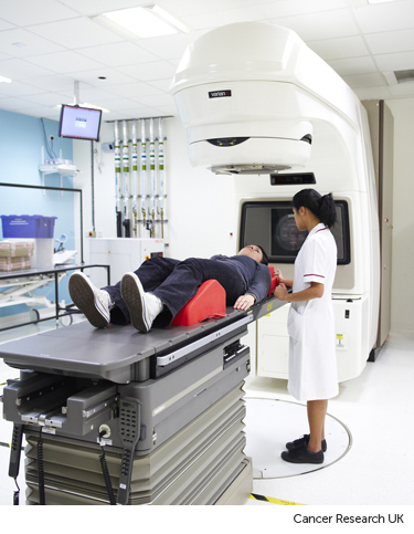
Stereotactic radiotherapy (SRT)
Stereotactic radiotherapy (SRT) gives radiotherapy from many different angles around the body. The beams meet at the tumour. This means the tumour receives a high dose of radiation and the tissues around it receive a much lower dose. This lowers the risk of side effects.
Usually you have between 1 and 8 treatments.
You might hear a few different terms for stereotactic radiotherapy. This can be confusing. Stereotactic treatment for the body might be called:
- stereotactic body radiotherapy (SBRT)
- stereotactic ablative radiotherapy (SABR)
Stereotactic radiotherapy to the brain might be called stereotactic radiosurgery (SRS). This is usually a single treatment. If you have more than one treatment to the brain, this is called stereotactic treatment.
When you might have stereotactic radiotherapy
This type of radiotherapy is mainly used to treat very small cancers, including:
- cancer in the lung
- cancer that started in the liver or cancer that has spread to the liver
- cancers in the lymph nodes
- spinal cord tumours
- cancer spread in the brain
Stereotactic radiotherapy can treat areas of the body that have had radiotherapy before. For example, if someone has had radiotherapy to their pelvis, they usually can't have radiotherapy to the same area again. But stereotactic treatment is so precise, it can often mean re-treatment is possible.
Research is being carried out to see what other cancers stereotactic treatment can help.
Planning stereotactic radiotherapy
Planning SRT involves several steps.
You start with having a CT scan in the radiotherapy department. You may also have MRI scans or PET scans of the area of your body to be treated. The information from these scans feeds directly into the radiotherapy planning computer.
The radiotherapy team then puts together a treatment plan.
Your doctor makes sure that all of the cancer is inside the radiotherapy field. And they avoid as much healthy tissue as possible. This reduces the risk of side effects.
Skin markings
The radiographers might make marks on your skin that act as reference points. This is to make sure every treatment session is accurate.
Radiotherapy masks and moulds
If you have radiotherapy to your head or neck, you may need to wear a radiotherapy mask during your treatment. This is also called a shell or mould.
Most types of masks are made of a mesh material with lots of small holes. You can have a mould for other parts of the body, such as the breast or limbs.
The mould or mask keeps the treatment area completely still. This is so your treatment will be as accurate as possible.
Metal markers
You may have small metal markers (fiducial markers) put in or near your tumour. This is common for prostate cancer.
To place the markers, your doctor inserts a needle into the area of skin over the cancer. They do this while you are having an ultrasound scan. Once the tip of the needle is in the right place your doctor releases small gold pellets or rods.
Your radiographers can then see the markers on x-rays and scans while you're having treatment. They act as a marker for the treatment area, and makes sure that the treatment is very accurate. Your radiographers will take x-rays or scans every day before treatment if you have this type of metal marker.
After the planning session
You usually have to wait a few days or up to 2 weeks for your treatment plan to be ready.
You then get an appointment for your first session of radiotherapy.
Having stereotactic radiotherapy
To have the treatment you lie on a radiotherapy couch. The radiographers help you to get into the right position and put on any moulds that you may need. They help make you as comfortable as possible.
Different machines can be used to give stereotactic radiotherapy. It is usually a machine called a Linear Accelerator (LINAC). But you might also have it on a CyberKnife machine.
Stereotactic radiotherapy with a LINAC

Once you are in the right position the radiographers leave the room. This is so they are not exposed to radiation.
The treatment lasts between 15 minutes to 2 hours, and sometimes longer. You might have the treatment in one go or it may be broken up with short breaks.
You will be alone, however your radiographers can see and hear you the whole time and can speak to you by intercom. It is important that you stay very still throughout the treatment.
You won't feel anything while you have the treatment and the machine doesn't touch you. The machine will beep from time to time.
Once the treatment is over your radiographers go back into the room. They help you get down from the treatment couch.
Stereotactic radiotherapy with a CyberKnife machine
The CyberKnife radiotherapy machine has a robotic arm. It moves around the treatment couch so it can give doses of radiation from different angles.
The radiotherapy machine usually takes x-rays every 10 to 20 seconds. The robotic arm uses the x-rays to correct its position. This allows your radiographers to target radiotherapy beams accurately at the tumour. Even if the tumour moves as you breathe.
Treatment may take between 30 minutes to 4 hours depending on the type of cancer and its position in the body. CyberKnife can treat multiple tumours at the same time. So it can be very useful for areas of cancer spread.
You may have the treatment in one go or it may be broken up with short breaks.
Side effects of stereotactic radiotherapy
Stereotactic radiotherapy targets the tumour very precisely. The risk of damage to normal surrounding tissues is low. Therefore the side effects may be less than with other types of radiotherapy.
Unfortunately you can still have side effects. This only affects the part of the body that the radiotherapy treatment is aimed at.
Getting stereotactic radiotherapy
Stereotactic radiotherapy is available at many treatment centres in the UK. This includes at NHS radiotherapy units that have specialised radiotherapy machines such as CyberKnife.
If you think this treatment may be right for your type of cancer ask your doctor. They can refer you to another centre if this would be an appropriate treatment for you.


