Types of surgery for eye cancer
Surgery for eye cancer is a very specialised operation. You might have an operation to remove certain types of eye cancer. For example, melanoma of the eye or lacrimal gland cancer. It is rare to have surgery for lymphoma of the eye.
Surgery might include:
- removing part of the eye
- removing the whole eye
- Mohs micrographic surgery
This section does not discuss all the details of these operations.
How you have surgery for eye cancer
Surgery for eye cancer is a specialised operation. You usually have surgery under general anaesthetic. So you will be asleep for the whole operation.
Before your surgery your ophthalmologist (eye surgeon) will explain the operation. They will discuss how it might affect you afterwards.
Your surgeon will preserve as much sight as possible. If your surgeon needs to remove your eye (an enucleation) then you will lose the sight in that eye.
Losing the sight in an eye can come as quite a shock. You will need time to come to terms with this change. Your healthcare team will help you. And there are organisations that can support you during this time. Ask questions as often as you need to, especially about your sight and appearance after the operation.
Removing part of the eye
Surgery to remove a cancer from the eyeball or around the eye is known as a tumour resection. The operation can also involve removing a small amount of healthy tissue around the cancer (a margin). This is to make sure all the cancer has been removed.
You may hear your specialist refer to this as eye sparing or eye conserving surgery. After your operation, you might have chemotherapy or radiotherapy or both.
The type of operation you have depends on where your cancer is. For example, you might need to have part of the choroid removed (a choroidectomy). Or, for small melanomas of the iris, you might have one of the following:
-
removal of part of the iris – iridectomy
-
removal of the iris and the ciliary body (the muscle that focuses the eye) - iridocyclectomy
Removing the whole eye
Your doctor may recommend surgery to remove the whole eye (enucleation). Or some people might have a bigger operation to remove the eye and surrounding tissue (orbital exenteration).
The operation to remove your eyeball is called an enucleation. This is often used for:
- eye melanomas too large for brachytherapy or proton beam radiotherapy
- a painful eye due to high pressure inside the eye
- a cancer that is growing through the wall of the eye
Your specialist will only suggest this if it is necessary. They will explain the operation and, if possible might show you pictures and photographs, so you know what to expect. This can help you prepare for how you might feel and look afterwards.
Your surgeon will remove your eyeball but leave your eyelids, brow and the surrounding skin in place.
During the operation to remove your eye, your surgeon fits a permanent eye implant into the socket. The implant is usually round like a ball and helps to fill some of the space where your eyeball once was.
The implant helps keep the structure of the eye socket and supports the artificial eye, which is fitted later. The implant is covered with eye tissue in the socket so you can’t see it. The area just appears pink. It is permanent and you can’t take it out.
Plastic implants have been used for decades and allow some eye movement. More recently new types have been used, an example is hydroxyapatite implants. They are made from an artificial form of sea coral. This material is very similar to bone in structure. Blood vessels can grow into it so it can work as part of the normal eye tissue.
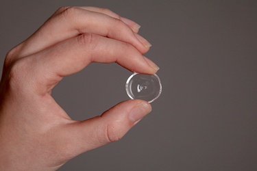
Your surgeon may also put a temporary plastic shell in the socket over the implant called a conformer. It is similar to a contact lens. The conformer helps to keep the shape of the eye socket while it heals. It has a hole in the middle that allows air to flow around the socket. It also helps to drain any build up of fluid.
The conformer is kept in for a few weeks. Before you go home you are shown how to remove and clean it. You may prefer to do this at your GP surgery, or at your hospital with a nurse until you are confident to do this on you own. After a few weeks the conformer is replaced with an artificial eye (prosthesis).

About 4 to 6 weeks after your surgery you have a temporary artificial eye fitted. At the same time you will be measured for an artificial eye made just for you.
The artificial eye is not round like a ball, but more like the shape of a big contact lens. It will have an eye painted onto it to match your remaining eye. It looks quite natural but you may not have the same the range of movement as the other eye.
You can take out your artificial eye whenever necessary.
Rarely a surgeon may recommend surgery to remove the eyeball and surrounding tissue. This includes the eyelid and the muscle and fat around the eye. This is a bigger operation and is called an orbital exenteration.
After surgery there is a wound around the eye socket. You usually have a dressing over the eye and this may be kept in place for about a week. The nurses will check and clean the area and show you how to look after it.
After a few months when the area has healed, a prosthesis is made to replace the part of the eye that was removed. This includes an artificial eye, eyelid and lashes.
It can be difficult to recover from and cope with this kind of surgery. Your healthcare team will support you throughout your recovery and follow up. You will also be offered counselling.
Mohs micrographic surgery
Mohs micrographic surgery is used to remove skin cancers. It aims to remove all the cancer, while sparing as much healthy tissue around it as possible. You may also hear this called Mohs surgical technique.
Your surgeon might use this surgery to remove skin cancers from delicate areas around your eye, such as the eyelid. For example squamous cell or basal cell skin cancers of the eye.
The surgeon removes the cancer with a very small margin around it and examines it under the microscope. If cancer is seen at the edge the surgeon removes a little more. They repeat this until they remove all the cancer.
The surgeon that does this operation is a specialist that is also trained in looking at specimens (pathologist).
Risks of surgery
All types of surgery have risks. Your surgeon will explain the risks to you in detail. These will include the risk of partial or total loss of eyesight due to various reasons, as well as infection and bleeding.
Ask your surgeon if you are unclear or concerned about any of the risks.
Removing part of the eye means there is a risk of cancer cells breaking away from the cancer and spreading to the surrounding tissue. This is rare and your surgeon will do all they can to reduce this risk.
Your nurse will monitor you regularly while you are in hospital. Before you go home, your team will go through what to look out for. And you should have a contact number to ring if you have any questions or concerns.
How you might feel after eye surgery
After eye surgery you can go through a range of emotions. It can take time to adjust and come to terms with how you feel, as well as managing practical changes.
Loss of sight in an eye can cause changes in depth perception. This is how you judge distance between things. In time you will get used to these differences. Your healthcare team will support you. And there are other organisations who can offer information and support.


